The protocol for Corneal Abrasions/Corneal Ulcers/Conjunctivitis:
Let’s break it down!

Within the medic’s scope of practice, there are only three ocular conditions that we’re looking for: Corneal Abrasion, Corneal Ulcer, and Conjunctivitis. Let’s talk a little bit about each:
Conjunctivitis: commonly known as “Pink eye”; it’s simply the inflammation of the conjunctiva (the transparent membranes lining the inside of the eyelid and the white of your eyes). Extremely common in kids and fairly common in adults, representing about 1% of all ER/Primary care visits. It can be caused by allergies, viruses, or bacteria.

Corneal Abrasion: a superficial scratch on the cornea (the “window” of the eye). Usually caused by foreign objects: dirt, dust, metal particles, contact lenses…even the edge of a piece of paper. Accounts for 80% of all visits for “eye trauma” in the ED. Very common in working-class men, specifically between the ages of 20-29 who work in the automobile industry. Naturally, it’s very common in soldiers as well.

Corneal Ulcer: Also known as “Keratitis”. It’s an open sore on the cornea that’s most commonly caused by some form of infection. It accounts for approximately 1 million ED and outpatient clinic visits annually and its incidence is increasing due to the increased use of contact lenses. Although not typically life-threatening, it is vision-threatening.


Corneal Abrasion: Corneal abrasions are very difficult to diagnose just by looking at it. Typically, you have to utilize fluorescein staining to identify the finer details of an abrasion. Any time somebody talks about any potential trauma to the eye or says something like “I feel something in my eye“, that’s your cue to pull out the fluorescein.

Corneal Ulcer: Easy one to diagnose. Although fluorescein staining can help identify the etiology of it (trauma that led to it), you can actually sometimes see it perfectly well with the naked eye. And of course, it will excruciatingly painful for the patient with the potential for vision loss. The video below show’s a patient with a real corneal ulcer emergency:
Conjunctivitis: Identifying conjunctivitis isn’t particularly difficult. Just looking for somebody with red, watery eyes. The tricky part is identifying what kind of conjunctivitis it is. The one we can treat is bacterial conjunctivitis; which is characterized by heavy drainage from the eye, typically on one side. Viral/allergic conjunctivitis is typically mild, low discharge, and affects both eyes.


Regardless of what eye condition you think it is, always perform an inspection. Below is two videos that show how to properly perform an eyelid inversion and a fluorescein stain:
If you suspect a foreign body that’s not immediately identifiable with the eyelid inversion, you can use saline to help flush it out. To do this, you can utilize an IV tubing, nasal cannula, etc. If you use a syringe, be careful not to squirt it too aggressively into the eye itself.

Corneal abrasions and mild cases of conjunctivitis will typically heal on their own w/o any intervention. The only two things we really care about are corneal ulcers and bacterial conjunctivitis.
Gatifloxacin is an antibiotic eye drop solution that just so happens to be the only ophthalmic solution within our scope. It’s a broad-spectrum antibiotic, which will help cover some of the common culprits: Staphylococcus aureus, Streptococcus viridans, and Streptococcus pneumonia. For treatment, you’ll provide them with the bottle and instruct them to take it qid (4X per day) or every 2hrs if it’s a corneal ulcer. Every day that they take antibiotics, they MUST come back for a fluorescein stain for a follow up to ensure that they are improving. Otherwise, oral antibiotics may be needed.

For pain, Tetracaine is our go-to. It’s basically lidocaine for the eye, which if you remember, is a local anesthetic; It numbs whatever it touches! It works within about 30 seconds, but it doesn’t last very long (only about 15 minutes). This can be a good thing to apply before you start poking around the eye or doing any procedure, but it’s not really a long-term pain management solution. You may be tempted to give the bottle of tetracaine to the patient for later use, but most texts don’t advocate for this due to it’s potential for toxicity if abused. Instead, you should utilize the Pain Management protocol and send the patient home with NSAIDs and/or Tylenol.


5. No patching
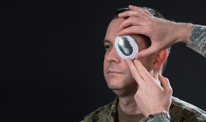
This is mostly a concern with severe ocular trauma, but in general, you should never “patch” or cover an eye with a loose substance. This will fail to provide any sort of protection to the patient’s eye. Instead, utilize a rigid eye shield or the patient eye pro if you believe that they’ll be in an environment that poses a risk to the already injured eye.
6. Reduce light exposure/stay indoors/wear sunglasses as feasible
Nothing remarkable here. Just helps protect the eye and facilitates healing
7. Monitor daily w/ fluorescein…
Taxing, but worth it. The patient should only need to be reevaluated and on antibiotic drops for a few days.

The prognosis for basic corneal abrasions is excellent, with most only requiring a few days t heal. If it is not improving, then we may have advanced etiology that requires at least a Routine evacuation. Due to the risk of vision loss, we should always provide a Priority evacuation for someone with a corneal ulcer. And although this protocol doesn’t necessarily address major trauma to the eye or LASIK flap dislocations, if you see something like the pictures below, the patient needs an Urgent evacuation!
Globe Rupture

LASIK flap dislocation

Good luck out there!
References
- EMRAP Corependium: Keratitis
- Conjunctivitis Epidemiology
- Advanced Tactical Paramedic Protocols Handbook. 10th ed., Breakaway Media LLC, 2016.