This is the protocol for Chest Pain:
Chest Pain
Let’s break it down!
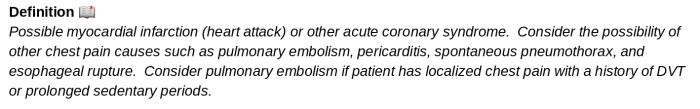
Chest pain is not a definitive diagnosis; it’s a chief complaint. It’s on us to figure out what exactly is causing it. The protocol lists several differentials for chest pain in the soldier population. Let’s briefly define what each of those are:
- Myocardial Infarction (“Heart Attack”): Refers to tissue damage inflicted on the heart muscles. Most commonly caused by atherosclerotic plaque build-up in the coronary arteries, but it can also occur from things like stimulant drug overdoses that cause severe constriction of the arteries. When tissue death is bad enough to cause ST elevation changes on an electrocardiogram (EKG), then it’s referred to as an ST-Elevation Myocardial Infarction or “STEMI“. Without ST-Elevation, it is referred to as a Non-ST-Elevation Myocardial Infarction or “NSTEMI“.

- Acute Coronary Syndrome (ACS): This is a catch-all phrase used to define any pathology that leads to a decreased blood flow to heart tissue. Myocardial Infarction is one of them, but ACS doesn’t always necessarily imply that the heart tissue is dying. Rather, it simply means that the heart is ischemic or oxygen-deprived from an acute reduction of blood flow. The 4 main types are:
-
- Stable Angina (transient cardiac ischemia w/o tissue death)
- Unstable Angina (persistent cardiac ischemia w/o tissue death)
- NSTEMI (non-ST elevation myocardial infarction)
- STEMI (ST-elevation myocardial infarction)
-

- Pericarditis: An inflammation of the pericardium, which is a fibrous sac-like membrane that surrounds the heart. Typically secondary to a viral infection unrelated to the heart.

- Spontaneous Pneumothorax: Refers to a pneumothorax that occurs suddenly in the absence of a traumatic mechanism or known lung disease. Often occurs through the bursting of “blebs” (small air-filled lesions in the lungs). Classically occurs in tall, young, slender male patients.
- Esophageal Rupture: It’s as scary as it sounds… a perforation of the esophagus. Usually occurs during endoscopy or other surgical procedures, but can also occur spontaneously after intense episodes of vomiting.
- Pulmonary Embolism (PE): A blockage or clot in a pulmonary artery. A history of deep vein thrombosis (DVT) is a major risk factor for pulmonary embolism. Also, prolonged sedentary periods (flights, surgeries, etc.) can cause clots to form, break off, and enter the pulmonary vasculature.


Despite our wide array of chest pain differentials, we’re only really looking for one in particular: Myocardial Infarction. In the clinical setting, heart attacks are both common and dangerous. Until proven otherwise, chest pain is always an MI.
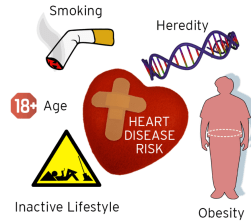
With that being said, every sign/symptom shown above is characterizing just that: a heart attack. When taking your SAMPLE HX, look for some of those things that indicate a risk for strained vessels and plaque build-up. In other words, look for someone who is old, fat, smokes a lot, and has a history of diabetes or heart disease. A combination of these risk factors would SCREAM acute coronary syndrome.
Each sign and symptom of a myocardial infarction can be explained as follows:
- Substernal pressure/chest pain: Classic symptom in response to added strain on the heart. Patients often say it feels like something is “sitting on my chest”
- Radiation to left arm/jaw: There are nerve connections between the left arm/jaw and the heart. When the nerves in the heart become irritated, a referred pain may occur in the left arm/jaw.
- Diaphoresis: Caused by a general sympathetic response to help perfuse the heart. Triggering the “fight or flight” response will always produce excessive sweating.
- Dyspnea: Increased ventilatory response in an effort to offset the cardiac ischemia and bring more oxygen to the heart.
If myocardial infarction progresses, the heart may ultimately fail to pump blood efficiently, leading to cardiogenic shock. As a result, fluid will back up into the pulmonary vasculature and cause bilateral rales/crackles. In addition, the patient may become hypertensive/hypotensive as the cardiogenic shock progresses.
Special note: These are all “classic” signs/symptoms of myocardial infarction. In other words, this is what’s common in the predominantly MALE soldier population. FEMALE soldiers often exhibit a different, and sometimes more subtle, set of symptoms 🕵🏼♀️ This can include nausea, fatigue, indigestion, etc.


The “MONA” treatment algorithm has been around for years. Although still considered to be generally effective as a temporizing treatment for myocardial infarctions, some special considerations exist for each drug:
Morphine Sulfate
Morphine is primarily used to treat the pain associated with infarction as opposed to fixing the infarction itself. Traditionally thought to have some cardioprotective benefits (decreased oxygen demand due to pain relief, etc.), but other studies have questioned its actual benefit. Regardless, this drug is generally safe to give.

Oxygen
It makes sense to provide oxygen to a patient who is struggling to provide adequate oxygenation to the heart. Definitely give it, but keep in mind that too much oxygen saturation (above 94%) has been shown to be harmful to patients experiencing MI’s due to the development of oxygen free radicals, which damage blood vessels.

Nitroglycerin
Nitroglycerin is a potent vasodilator. It helps dilate the clogged coronary vessels to restore adequate perfusion. However, nitroglycerin can be harmful to patients experiencing infarctions on the inferior portion of the heart due to its detrimental effect on cardiac preload. In the absence of 12-lead EKG capabilities, use it with caution.

Aspirin
Contrary to popular belief, the plaque build-up in coronary arteries by itself isn’t typically what causes myocardial infarctions. Rather, it’s the inflammatory platelet response that occurs after the plaque ruptures. The platelets aggressively bind to the plaque site, causing rapid occlusion of the vessel. Aspirin helps prevent this by making the platelets “slippery“. In other words, it keeps the platelets from being able to bind with( each other. Out of all of the drugs in the MONA pneumonic, this is by far the most life-saving (reduces vascular death by 21%)


2. Establish IV access
Although not typically needed for pre-hospital treatment, it’s a must-have for continued care. Later, the patient may be given additional IV medications, including thrombolytics (clot-busting agents)
3. Avoid all exertion
Engaging in physical activity will increase the myocardial oxygen demand of the heart, which is counter productive when heart tissue is actively dying from ischemia. This is obvious enough, but be particularly careful even while moving the patient from one place to another. Simply walking to the ambulance or going down a flight of stairs can be enough to make your patient crash.
4. Pulse oximetry and cardiac monitor (if available)
Serial Spo2 and EKG checks can help identify patient improvement or decompensation. Bad signs would include a drop in Sp02 (below 94%) or further ST-elevation in EKG leads, both indicating a decrease in myocardial perfusion.

DON’T MESS WITH CHEST PAIN. Unless you’re sure it’s something benign like costochondritis or anxiety, you’re always going to give these patients an Urgent evacuation. Definitive care for a myocardial infarction will likely require a cath lab for clot removal or reperfusion procedures. If you suspect one of the other differentials we discussed (esophageal rupture, PE, etc.), the evacuation criteria will still be the same.
In addition, the patient should be transferred over to medics or providers with Advanced Cardiac Life Support (ACLS) capabilities, if possible. With the right equipment, these personnel will be able to perform 12-lead EKGs, confirm the diagnosis, and be better prepared in the event that the patient decompensates into cardiogenic shock or cardiac arrest.
Till next time!
References
- EMRAP Corependium: Acute Coronary Syndrome
- Uptodate: Overview of the acute management of myocardial infarction
- Stapczynski JS, Tintinalli JE. Tintinallis Emergency Medicine: a Comprehensive Study Guide, 8th Edition. New York: McGraw-Hill Education; 2016.
- Advanced Tactical Paramedic Protocols Handbook. 10th ed., Breakaway Media LLC, 2016.